Thành viên:Naazulene/Dây thần kinh gai
| Spinal nerve | |
|---|---|

| |
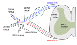
| The formation of the spinal nerve from the posterior and anterior roots | |
| Latinh | nervus spinalis |
Dây thần kinh gai là các bó neuron mọc ra từ tủy gai trong xương sống. Các dây này là là dây thần kinh hỗn hợp, dẫn truyền cả tín hiệu vận động và cảm giác. Cơ thể người có 31 đôi dây thần kinh gai (mỗi cặp có dây bên trái và bên phải).[1][2]Dựa trên các vùng của cột sống, 31 dây đó được phân thành các vùng: cổ (8), ngực (12), thắt lưng (5), cùng (5), cụt (1).[1] Các dây thần kinh gai là thành phần của hệ thần kinh ngoại biên, cùng với dây thần kinh sọ.[1]
Từ nguyên
sửaChữ gai trong dây thần kinh gai hay tủy gai không thể hiện hình thể của chúng, mà nó có nghĩa "liên quan đến cột sống", được dịch từ chữ épinière (tiếng Pháp). Épinière có nguồn gốc từ chữ spina (tiếng Latin), nghĩa là cái gai trên cây.[3] Cột sống của người có hình thể ngoài giống thân cây có gai.
Cấu trúc
sửaMỗi dây thần kinh gai là một dây thần kinh hỗn hợp, hình thành từ các sợi thần kinh rễ từ rễ bụng và rễ lưng. Rễ lưng còn được gọi là rễ sau, rễ cảm giác; nó mang tín hiệu cảm giác từ cơ quan về tủy sống và não. Rễ bụng còn gọi là rễ trước hay rễ vận động, nó mang tín hiệu vận động từ hệ thần kinh trung ương ra các cơ quan. Mỗi rễ này tỉa ra từ tủy gai, luồn qua các lỗ ghép (lỗ gian đốt sống) giữa mỗi hai đốt sống, kết hợp để tạo thành dây thần kinh hỗn hợp, rồi đi đến các cơ quan. Các đôi dây thần kinh gai chi phối các vùng từ cổ trở xuống. Các dây thần kinh gai chia thành nhánh trước và nhánh sau (hoặc nhánh bụng và nhánh lưng, mỗi nhánh đều là dây hỗn hợp), các nhánh trước thường kết hợp với nhau để tạo thành các đám rối.
Về tên gọi, ở vùng ngực (T1 - T12) và thắt lưng (L1 - L5), đôi dây thần kinh mang tên của đốt sống ngay trên nó. Ở vùng cổ, đôi dây thần kinh mang tên của đốt sống dưới nó (C1 - C7), còn C8 là đôi dây ở trên đốt sống T1. Ở vùng cùng, các đôi dây thần kinh luồn qua các lỗ trên xương cùng thay vì luồn qua lỗ gian đốt sống và được đánh số từ trên xuống dưới (S1 - S5). Có 1 đôi dây thần kinh cụt (Co).
Các nhánh lưng chi phối vùng sau của thân mình. Các nhánh bụng chi phối vùng trước của thân mình và các chi. Ngoài ra còn có một số nhánh màng não luồn ngược về lỗ ghép để chi phối cột sống (các dây chằng, màng cứng, mạch máu, đĩa gian đốt sống, khớp mặt và màng quanh xương của đốt sống). Mỗi nhánh đều có dây thần kinh cảm giác và dây thần kinh vận động.
1. Somatic efferent.
2. Somatic afferent.
3,4,5. Sympathetic efferent.
6,7. Autonomic afferent.
Các nhánh bụng thường ghép lại với nhau để hình thành các đám rối thần kinh. Các dây trong đám rối thần kinh đi cùng nhau đến cơ quan đích. Những đám rối thần kinh lớn là đám rối cổ, đám rối cánh tay, đám rối thắt lưng, đám rối cùng và đám rối cụt (đám rối cụt nhỏ hơn nhiều).[4]
Với các nhánh trước, từ khi ra khỏi xương sống, các đoạn của dây thần kinh được đặt tên kiểu khác, theo thứ tự từ trong ra là: rễ, thân, ngành, bó, nhánh tận (tiếng Anh: root, trunk, division, cord, branch). Trong đó các rễ tương ứng với nhánh trước, và rễ này cũng tách bạch khỏi rễ bụng và rễ lưng. Vì các dây thần kinh có thể dung hợp hoặc tách ra, số lượng của các đơn vị này trong cùng đám rối có thể khác nhau (ví dụ: đám rối cánh tay có 5 rễ, 3 thân, 6 ngành, 3 bó, và nhiều nhánh tận). Trong tiếng Việt, các nhánh tận được đặt tên theo cấu trúc thần kinh [từ mô tả], ví dụ như thần kinh cơ bì.
Thần kinh định khu
sửaCác dây cổ
sửaCác dây thần kinh cổ là các dây thần kinh phát ra từ tủy gai trong các đốt sống cổ. Dù chỉ có 7 đốt sống cổ, có tận 8 đôi dây thần kinh cổ. Dây thần kinh C1–C7 luồn ra trên đốt sống có số tương ứng, còn C8 luồn dưới đốt sống C8. Các đôi dây thần kinh khác đều luồn dưới đốt sống cùng số.
Phân bố sau bao gồm dây thần kinh dưới chẩm (C1), dây thần kinh chẩm to (C2) và dây thần kinh chẩm thứ ba (C3). Phân bố trước bao gồm đám rối cổ (bốn dây đầu) và đám rối cánh tay (bốn dây sau, T1).
Các nhánh trước (rễ) của bốn dây cổ đầu nối với nhau thành 3 quai tạo thành đám rối cổ. Đám rối cổ phân thành ba loại nhánh:[5]
- Cảm giác: dây tai lớn, dây chẩm nhỏ, dây ngang cổ và dây trên đòn
- Vận động: chi phôi các cơ vùng gáy và cổ
- Nối: Dây thần kinh cổ C1 - C3 nối với dây thần kinh XII tạo một vòng dây thần kinh gọi là quai cổ. Vòng này chi phối các cơ ức móng, cơ ức giáp và cơ vai móng.
Các nhánh trước (rễ) của bốn dây cổ sau và dây T1 tạo thành đám rối cánh tay (5 rễ, 3 thân, 6 ngành (3 ngành trước và 3 ngành sau), 3 bó) từ đó cho ra 7 nhánh tận chi phối chi trên. Từ đám rối cánh tay, quan trọng nhất là 5 dây thần kinh này:[5]
- Thần kinh cơ bì
- Thần kinh nách
- Thần kinh quay
- Thần kinh giữa
- Thần kinh trụ
Các dây ngực
sửaCó 12 đôi dây thần kinh ngực luồn ra từ các đốt sống ngực. Mỗi dây thần kinh ngực T1-T12 luồn ra từ dưới đốt sống cùng số. Một vài nhánh đi trực tiếp đến hách bên cột sống để tham gia chi phối các cơ quan là tuyến của đầu, cổ, ngực và bụng.
Các dây ngực không tạo thành đám rối mà chủ yếu tạo thành các dây gian sườn có chức năng chi phối vận động cho các cơ gian sườn và chi phối cảm giác cho thành ngực và thành bụng trước trên.
Phân bố trước
sửaCác dây gian sườn đến từ các dây T1-T11, chạy giữa các xương sườn. Ở T2 và T3, cách nhánh xa hình thành nên dây thần kinh gian ngực cánh tay. Đây thần kình dưới ngực đến từ dây T12, và chạy dưới xương sườn số 12.
Posterior divisions
sửaThe medial branches (ramus medialis) of the posterior branches of the upper six thoracic nerves run between the semispinalis dorsi and multifidus, which they supply; they then pierce the rhomboid and trapezius muscles, and reach the skin by the sides of the spinous processes. This sensitive branch is called the medial cutaneous ramus.
The medial branches of the lower six are distributed chiefly to the multifidus and longissimus dorsi, occasionally they give off filaments to the skin near the middle line. This sensitive branch is called the posterior cutaneous ramus.
Lumbar nerves
sửaThe lumbar nerves are the five spinal nerves emerging from the lumbar vertebrae. They are divided into posterior and anterior divisions.
Posterior divisions
sửaThe medial branches of the posterior divisions of the lumbar nerves run close to the articular processes of the vertebrae and end in the multifidus muscle.
The laterals supply the erector spinae muscles.
The upper three give off cutaneous nerves which pierce the aponeurosis of the latissimus dorsi at the lateral border of the erector spinae muscles, and descend across the posterior part of the iliac crest to the skin of the buttock, some of their twigs running as far as the level of the greater trochanter.
Anterior divisions
sửaThe anterior divisions of the lumbar nerves (rami anteriores) increase in size from above downward. They are joined, near their origins, by gray rami communicantes from the lumbar ganglia of the sympathetic trunk. These rami consist of long, slender branches which accompany the lumbar arteries around the sides of the vertebral bodies, beneath the psoas major. Their arrangement is somewhat irregular: one ganglion may give rami to two lumbar nerves, or one lumbar nerve may receive rami from two ganglia.
The first and second, and sometimes the third and fourth lumbar nerves are each connected with the lumbar part of the sympathetic trunk by a white ramus communicans.
The nerves pass obliquely outward behind the psoas major, or between its fasciculi, distributing filaments to it and the quadratus lumborum.
The first three and the greater part of the fourth are connected together in this situation by anastomotic loops, and form the lumbar plexus.
The smaller part of the fourth joins with the fifth to form the lumbosacral trunk, which assists in the formation of the sacral plexus. The fourth nerve is named the furcal nerve, from the fact that it is subdivided between the two plexuses.
Sacral nerves
sửaThe sacral nerves are the five pairs of spinal nerves which exit the sacrum at the lower end of the vertebral column. The roots of these nerves begin inside the vertebral column at the level of the L1 vertebra, where the cauda equina begins, and then descend into the sacrum.[6][7]
There are five paired sacral nerves, half of them arising through the sacrum on the left side and the other half on the right side. Each nerve emerges in two divisions: one division through the anterior sacral foramina and the other division through the posterior sacral foramina.[6]
The nerves divide into branches and the branches from different nerves join with one another, some of them also joining with lumbar or coccygeal nerve branches. These anastomoses of nerves form the sacral plexus and the lumbosacral plexus. The branches of these plexus give rise to nerves that supply much of the hip, thigh, leg and foot.[6][8]
The sacral nerves have both afferent and efferent fibers, thus they are responsible for part of the sensory perception and the movements of the lower extremities of the human body. From the S2, S3 and S4 arise the pudendal nerve and parasympathetic fibers whose electrical potential supply the descending colon and rectum, urinary bladder and genital organs. These pathways have both afferent and efferent fibers and, this way, they are responsible for conduction of sensory information from these pelvic organs to the central nervous system (CNS) and motor impulses from the CNS to the pelvis that control the movements of these pelvic organs.[8]
Coccygeal nerves
sửaThe bilateral coccygeal nerves, Co, are the 31st pair of spinal nerves. It arises from the conus medullaris, and its ventral ramus helps form the coccygeal plexus. It does not divide into a medial and lateral branch. Its fibers are distributed to the skin superficial and posterior to the coccyx bone via the anococcygeal nerve of the coccygeal nerve plexus.
Function
sửa| Số | Chức năng vận động |
|---|---|
| C1-C6 | Các cơ gấp cổ |
| C1-T1 | Các cơ duỗi cổ |
| C3-C5 | Cơ hoành (chủ yếu là C4) |
| C5, C6 | Cử động vai, cử động cơ thang, |
Spinal plexuses
sửaA spinal plexus is a weblike nerve plexus formed by the anterior nerve roots that branch and merge repeatedly. The only region that does not have a plexus is the thoracic region. The small cervical plexus is in the neck, the brachial plexus is in the shoulder, the lumbar plexus is in the lower back, beneath this is the sacral plexus, and next to the lower sacrum and coccyx is the very small coccygeal plexus.[4]
Clinical significance
sửaThe muscles that one particular spinal root supplies are that nerve's myotome, and the dermatomes are the areas of sensory innervation on the skin for each spinal nerve. Lesions of one or more nerve roots result in typical patterns of neurologic defects (muscle weakness, abnormal sensation, changes in reflexes) that allow localization of the responsible lesion.
There are several procedures used in sacral nerve stimulation for the treatment of various related disorders.
Sciatica is generally caused by the compression of lumbar nerves L4, or L5 or sacral nerves S1, S2, or S3, or by compression of the sciatic nerve itself
Additional Images
sửa-
A portion of the spinal cord, showing its right lateral surface. The dura is opened and arranged to show the nerve roots.
-
Distribution of the cutaneous nerves. Ventral aspect.
-
Distribution of the cutaneous nerves. Dorsal aspect.
-
The spinal cord with dura cut open, showing the exits of the spinal nerves.
-
The spinal cord showing how the anterior and posterior roots join in the spinal nerves.
-
A longer view of the spinal cord.
-
Projections of the spinal cord into the nerves (red motor, blue sensory).
-
Projections of the spinal cord into the nerves (red motor, blue sensory).
-
Schematic diagram of cervical plexus.
- Dissection images
-
Cerebrum. Inferior view. Deep dissection.
-
Cerebrum. Inferior view. Deep dissection.
-
Spinal nerves. Spinal cord and vertebral canal. Deep dissection.
See also
sửaReferences
sửa- ^ a b c Kaiser, JT; Lugo-Pico, JG (tháng 1 năm 2024). Neuroanatomy, Spinal Nerves. PMID 31194375.
- ^ “A Neurosurgeon's Overview of the Anatomy of the Spine and Peripheral Nervous System”. www.aans.org (bằng tiếng Anh). Truy cập ngày 21 tháng 12 năm 2023.
- ^ “spine | Etymology of spine by etymonline”. www.etymonline.com (bằng tiếng Anh). Truy cập ngày 4 tháng 10 năm 2024.
- ^ a b Saladin, Kenneth S. (2011). Human anatomy (ấn bản thứ 3). New York: McGraw-Hill. tr. 382–388. ISBN 9780071222075.
- ^ a b PGS.TS. Phạm Đăng Điệu. Giản Yếu Giải Phẫu Người. ISBN 978-604-66-6309-6.
- ^ a b c 1. Anatomy, descriptive and surgical: Gray's anatomy. Gray, Henry. Philadelphia : Courage Books/Running Press, 1974
- ^ 2. Clinically Oriented Anatomy. Moore, Keith L. Philadelphia : Wolters Kluwer Health/Lippincott Williams & Wilkins, 2010 (6th ed)
- ^ a b 3. Human Neuroanatomy. Carpenter, Malcolm B. Baltimore : Williams & Wilkins Co., 1976 (7th ed)
- Blumenfeld H. 'Neuroanatomy Through Clinical Cases'. Sunderland, Mass: Sinauer Associates; 2002.
- Drake RL, Vogl W, Mitchell AWM. 'Gray's Anatomy for Students'. New York: Elsevier; 2005:69-70.
- Ropper AH, Samuels MA. 'Adams and Victor's Principles of Neurology'. Ninth Edition. New York: McGraw Hill; 2009.